What Is the Cervical DNA Dtex Test and How Does It Work?
The Cervical DNA Dtex Test is an advanced genomic screening tool designed to detect early signs of cervical cancer. It uses FISH technology (Fluorescence In Situ Hybridization) to identify irreversible DNA damage caused by high risk HPV infections often before visible cellular changes occur. By analyzing cervical cells at the molecular level, the test helps doctors assess a woman’s real risk of developing precancerous or cancerous lesions, enabling earlier and more precise medical decisions.

Understanding FISH Technology and Its Role in Early Detection of Cervical Cancer
Cervical cancer remains one of the most preventable yet deadly forms of cancer in women especially when detected too late. Traditional methods like the Pap smear and HPV tests are useful but can miss early molecular changes in cervical cells. That’s where the Cervical DNA Dtex Test comes in a breakthrough in early cancer detection that uses cutting-edge FISH technology to analyze DNA damage at the cellular level.
What is FISH Technology?
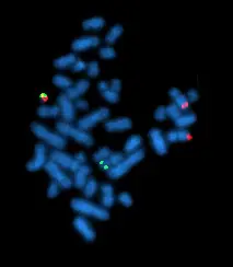
FISH, or Fluorescence In Situ Hybridization, is an advanced molecular cytogenetic technique used to visualize specific DNA sequences inside cells. It employs fluorescently labeled DNA probes that hybridize to complementary sequences on chromosomes or within the cell nucleus. When viewed under a fluorescence microscope, these probes light up, revealing detailed information about the genetic material in each cell.
Unlike traditional tests that rely on changes in cell shape or the presence of viruses, FISH directly detects genetic abnormalities at the DNA leveloffering a more precise snapshot of the cell’s health.
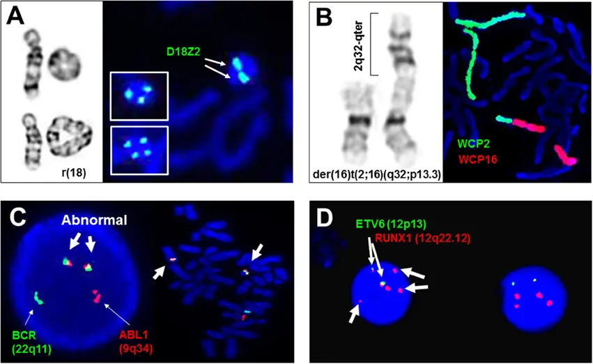
Adjunctive and diagnostic assays of FISH in clinical cytogenetics
How Does FISH Work in Cervical Cancer Detection?
In the context of cervical cancer, FISH technology is used to identify irreversible DNA damage caused by infection with high-risk of HPV. Here’s how it functions:
- Targeted Probes: FISH probes are designed to bind to specific regions of the genome that are frequently altered when HPV triggers malignant transformation. These include chromosomal regions involved in cell cycle regulation and tumor suppression.
- Detection of Genetic Changes: The fluorescent signals show whether those key regions are intact, duplicated, missing, or rearranged. Patterns of abnormal signal numbers or distributions indicate genomic instability—a hallmark of precancerous or cancerous cells.
- Early Warning Signs: Since DNA damage occurs before cells visibly change shape or become cancerous, FISH can detect precancerous lesions at a very early stage, sometimes even before abnormalities appear in a Pap smear.
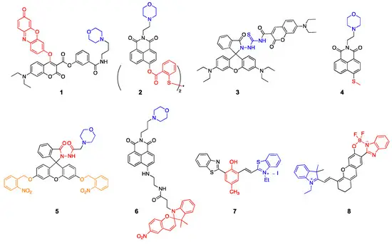
Lysosome-targeted fluorescent probes (blue: targeting moiety; red: responsive moiety; orange: photocaging group).
Advantages of FISH in Early Detection
- Sensitivity: FISH can reveal subtle genetic changes that other tests miss.
- Specificity: It helps differentiate between transient HPV infections and those causing dangerous cellular changes.
- Risk Stratification: Clinicians can better identify which patients are at high risk and require immediate follow-up or treatment.
- Reduced Over-Treatment: By targeting only those with confirmed DNA damage, unnecessary invasive procedures like biopsies or treatments can be avoided.
Real-World Impact
Early detection using FISH technology means more effective interventions, improved patient outcomes, and a reduction in cervical cancer rates. For women with ambiguous Pap or HPV results, the Cervical DNA Dtex Test based on FISH offers clarity and peace of mind.